Hantavirus Symptoms, Transmission, and Risk Factors
Most hantavirus cases go undiagnosed because mild infections never reach medical attention.
Global estimates place annual infections somewhere between 10,000 and more than 100,000 cases. That ten-fold spread is not imprecision in the counting. It reflects how many infections, particularly mild or asymptomatic HFRS cases that resolve before anyone orders a serology panel, never enter any registry. Serological surveys consistently show that confirmed case counts understate true incidence by a meaningful margin, and the gap between what circulates and what gets counted is wide enough to distort how seriously most clinicians are trained to take this pathogen. But what if the numbers we rely on are themselves part of the problem — not wrong exactly, but systematically incomplete in ways that shape clinical instincts in the wrong direction?
The largest burden sits in Asia and Europe. China and South Korea account for many thousands of cases annually. Europe recorded just under 1,900 cases across all hantavirus strains in 2023. None of this registers prominently in American public health discourse, partly because HPS dominates U.S. coverage and partly because absolute numbers in the Americas are smaller. Across eight countries in the Americas, 299 documented cases and 59 deaths were reported in 2025. The U.S. contribution to that total, drawn from surveillance data compiled since 1993 through the end of 2023, is 890 total confirmed cases, 35% of which resulted in death. In 2024 the U.S. recorded 20 cases; in 2025, 40 cases; between January 1 and May 9, 2026, four additional cases had been documented.
Those raw counts make hantavirus sound negligible, and for years that is more or less how it was treated in settings outside the Four Corners region. A disease that kills roughly one in three confirmed patients commands attention disproportionate to its absolute incidence, particularly when 94% of U.S. cases have occurred west of the Mississippi River. That geographic concentration is not reassuring; it is clarifying. Rarity and lethality sit in genuine tension here, and it is that tension, not either figure alone, that warrants careful attention.
How the Virus Moves from Rodent to Human
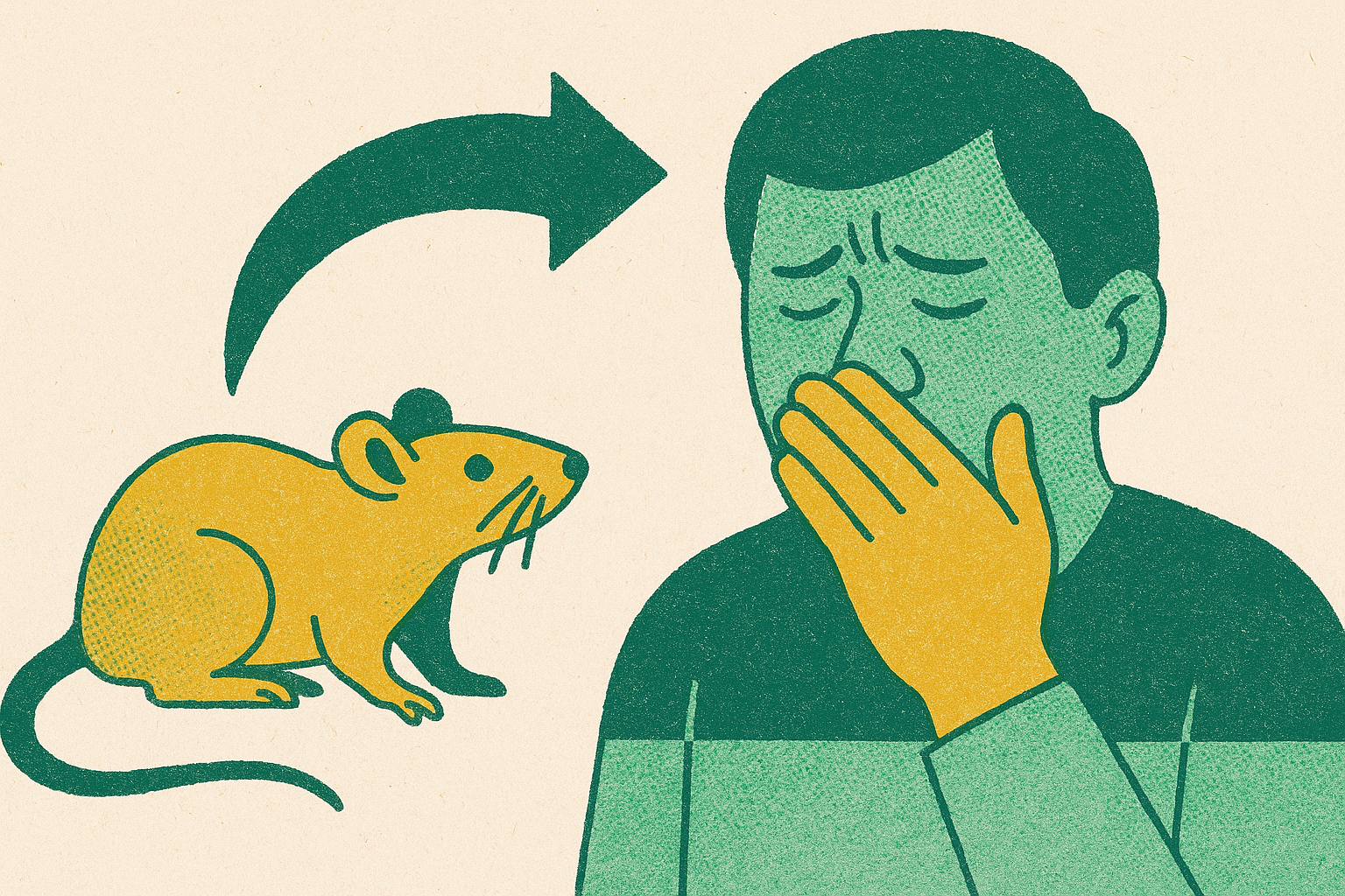
The dominant transmission route is inhalation of aerosolized particles from infected rodent urine, droppings, or saliva. Secondary routes exist: touching the mouth or nose after handling contaminated material, or, rarely, a bite. But the aerosol pathway shapes nearly all prevention logic, because disturbance, not mere presence, is what converts a contaminated space into a dangerous one. Sweeping or vacuuming launches particles into breathable air that would otherwise stay settled. That single distinction drives most of what matters in prevention.
Hantavirus survives outside the host for probably less than one week indoors under typical conditions, considerably less when exposed to direct sunlight. A recently vacated but sealed space carries more risk than one aired out for several days, and "recently" here means days, not hours. That temporal window matters when assessing an old cabin or a shed sealed through winter, and it is the kind of detail that tends not to surface until after someone is already ill.
In North America, the primary reservoir species are the deer mouse, white-footed mouse, cotton rat, and rice rat. An estimated 15% of deer mice carry Sin Nombre virus. The house mouse and Norway rat, the species most commonly encountered in urban environments, are not known carriers. Urban and rural populations face genuinely distinct exposure profiles, a distinction that gets flattened whenever hantavirus appears in generalist public health materials.
Insects do not transmit hantavirus. Ticks, fleas, and mosquitoes are non-vectors, despite the intuitive assumption that a rodent-borne pathogen might travel through the same channels as Lyme disease or plague. Human-to-human transmission has been documented only for Andes virus in South America, the reservoir of which is the long-tailed pygmy rice rat, and that transmission appears to require close, prolonged contact during early illness among household members or intimate partners. No other hantavirus strain has demonstrated person-to-person spread. For all practical purposes outside South America, this virus does not pass between people.
Who Faces the Highest Exposure Risk and Why
The intuition that hantavirus is an exotic wilderness disease does not survive contact with surveillance data. Approximately 70% of cases are associated with peridomestic exposure, meaning contact in or immediately around the home. Rodents reach where people live. At the same time, 93% of cases in Americas surveillance involved rural settings, so the risk is concentrated without being exotic. Rural proximity to rodent habitat is the operative variable, not remoteness for its own sake.
High-risk activities cluster around disturbance of enclosed or long-undisturbed spaces: cleaning rodent droppings or nests without respiratory protection; opening cabins, sheds, barns, storage buildings, garages, crawl spaces, or attics sealed for weeks or months; camping in trail shelters where deer mice have likely nested. One underappreciated exposure route is vehicle storage. Deer mice nest in cabin air filters, ductwork, and ventilation systems, so the first time a stored vehicle runs its HVAC, the driver may be inhaling aerosolized material before a single window has been cracked.
Occupational risk groups include farmers, ranchers, forestry workers, rural construction workers, pest control technicians, and wildlife biologists conducting field research. Regular entry into undisturbed structures or active rodent habitat is the common thread.
Geographically, Arizona led U.S. case counts over the most recent five-year reporting period with 26 cases, followed by New Mexico at 25 and Colorado at 13. The Four Corners region has retained its status as the highest-burden zone since the 1993 outbreak. Rodent population cycles compound this further: in years following mild winters or abundant food supply, deer mouse populations expand, and with them the probability that any given enclosed space has been colonized. Rural setting, enclosed and disturbed space, western geography, elevated post-boom rodent density. These four variables together give a clinician, or a careful layperson, a workable framework for evaluating exposure risk before it becomes clinical.
The Prodromal Phase: Why Early Symptoms Are So Easy to Dismiss
The incubation period runs from one to eight weeks after exposure. That range is long enough to sever the cognitive link between a specific rodent encounter and eventual illness onset. By the time symptoms appear, many patients have forgotten the exposure entirely, or dismissed it as inconsequential in the moment, which is a perfectly reasonable thing to do when you are cleaning out a garage on a Saturday afternoon and feel fine by Sunday.
When symptoms arrive, they come in a prodromal phase lasting roughly one to five days. Pan-American Health Organization clinical data from 43 cases with documented symptom profiles show fever in 93% of patients, headache in 84%, arthralgias in 72%, and myalgia in 65%. Those four findings describe dozens of common infections. Flu is the immediate differential, and an understandable one. What complicates triage is that dyspnea appeared in 60% of those same cases and tachypnea in 49%. Shortness of breath this early in an illness course is unusual for influenza. In a patient with a plausible rodent exposure history, respiratory symptoms appearing alongside fever and severe myalgia should raise clinical suspicion immediately, not after the chest film comes back.
One laboratory finding distinguishes hantavirus from common flu at this stage: thrombocytopenia, a falling platelet count. Patients do not feel it happening. But if blood work is obtained, thrombocytopenia in the context of fever, myalgia, and relevant exposure history should prompt a hantavirus workup without delay. That raises an important question: if this diagnostic signal is available, why does the prodromal window remain the interval in which the diagnosis is most reliably missed? The answer is not ignorance of the finding — it is that the workup is rarely ordered unless someone has already thought of hantavirus, and the default assumption pushes strongly toward flu. The prodromal phase is the only interval in which the trajectory of care can be meaningfully altered, and that is precisely why closing that diagnostic gap matters.
The Cardiopulmonary Phase in HPS and the Renal Crisis in HFRS
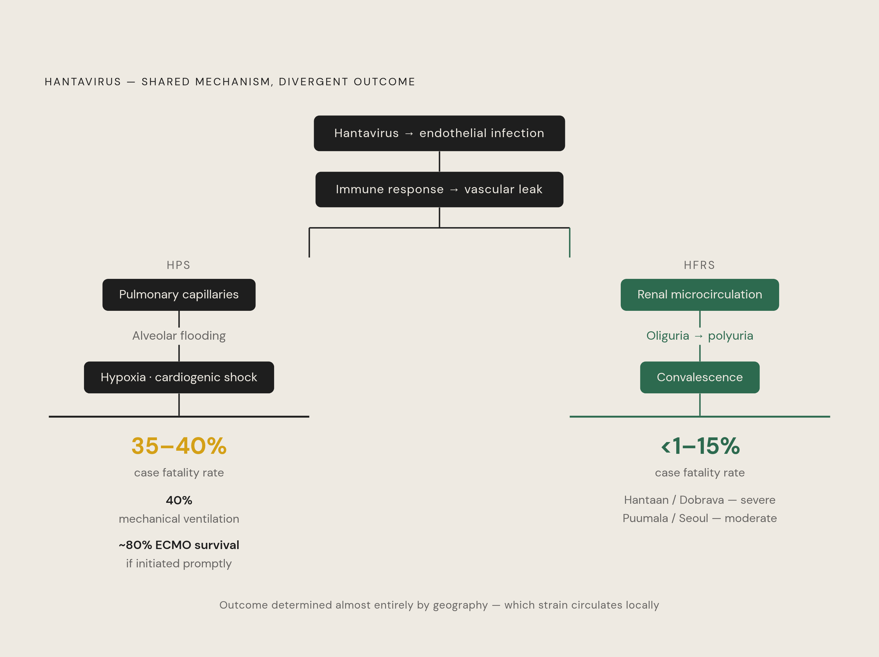
Both syndromes share a mechanism: vascular injury driven by the immune response, not direct viral cytotoxicity. Hantaviruses infect the endothelial cells lining small blood vessels, and what follows is the body's defenses damaging itself. The virus is not destroying tissue directly; it is redirecting the immune response into doing so. How quickly and severely that unfolds reflects the vigor of that response, which creates an uncomfortable clinical irony: robust immune function offers no straightforward protection and may actually accelerate the cascade.
In HPS, the cardiopulmonary phase begins within roughly ten days of illness onset. Pulmonary capillary leakage floods the alveoli with plasma, producing edema and progressive hypoxia. Tachycardia, arrhythmias, and cardiogenic shock follow as the heart fails to maintain output against a collapsing volume state. Approximately 40% of HPS cases require mechanical ventilation. The case fatality rate for HPS in the Americas runs between 35% and 40%. At the University of New Mexico, early extracorporeal membrane oxygenation (ECMO) has demonstrated roughly 70% success when initiated promptly, with survival approaching 80% in that setting. The same disease, recognized at different moments in its course, produces very different outcomes.
HFRS follows the same immune-driven vascular leak mechanism but targets the renal microcirculation rather than the pulmonary capillaries. Kidney involvement is universal, progressing through oliguria, then polyuria, then gradual convalescence. Hemorrhagic manifestations range from cutaneous petechiae to severe internal bleeding; in the gravest cases, disseminated intravascular coagulation develops and is considered a primary cause of death. Strain determines severity in ways that are clinically consequential: Hantaan and Dobrava viruses produce severe HFRS, while Seoul, Saaremaa, and Puumala generally cause more moderate disease. Case fatality rates for HFRS range from less than 1% to approximately 15% depending on strain, considerably lower than HPS but not benign at the upper end.
Same immunopathogenic mechanism, different anatomical target, fatality rates that diverge sharply between syndromes. Which crisis a given patient faces is largely determined by geography.
Recovery and What Persists After the Acute Phase
HPS recovery is slow. The convalescent phase typically extends across months, and breathing difficulties can persist for up to two years in some patients. There is no approved antiviral treatment. Management is entirely supportive: supplemental oxygen, mechanical ventilation, ECMO in severe cases. Clinical skill and attentiveness matter enormously within that constraint, but the constraint itself is real. There is no drug that reverses the cardiopulmonary cascade once it is fully established.
HFRS recovery follows a more staged course, with oliguria giving way to polyuria as renal function returns, then gradual normalization over weeks to months. Most patients with moderate-strain HFRS recover kidney function, though monitoring during convalescence remains necessary to catch cases where recovery stalls.
The 35% death rate in U.S. surveillance and the 22.9% case lethality reported in Paraguay in 2025 (a figure exceeding that country's four-year average of 15.9%) are outcomes that accumulate in a therapeutic landscape where intervention arrives too late once the crisis is fully declared. But how does this affect our original promise of meaningful intervention? It sharpens it: what can actually be shaped sits earlier — exposure prevention, and the prodromal window when early suspicion can still drive supportive care before hemodynamic collapse.
Reducing Exposure Risk Through Targeted Precautions
Prevention logic follows directly from transmission biology. If the primary route is aerosol inhalation during disturbance of contaminated spaces, protection means either eliminating the disturbance or controlling what is inhaled during it. Neither approach requires professional intervention in most situations.
Before entering a long-closed space, whether a cabin sealed through winter, a storage shed, a barn, or a crawl space, air it out for at least 30 minutes. Do not sweep or vacuum rodent droppings dry. Wet-wipe or spray contaminated surfaces with disinfectant first to suppress aerosolization, then remove the material. Use an N95 respirator, not a standard dust mask; the particle sizes at issue require a rated filter. Wear rubber or latex gloves and dispose of or disinfect them after use.
Around the home and property, seal gaps around pipes, foundations, and utility lines. Store food, including pet food and birdseed, in rodent-proof containers. Keep woodpiles, brush piles, and debris away from the structure itself, since these create nesting habitat adjacent to living spaces. If a vehicle has been stored outdoors or in a garage for any meaningful period, inspect and replace the cabin air filter before operating the HVAC system with occupants inside.
For camping and outdoor recreation, sleep in a tent with a sealed floor rather than on open ground, and inspect enclosed trail shelters before settling in. In occupational contexts, workers in agriculture, forestry, pest control, and wildlife research should follow site-specific respiratory protection protocols consistent with CDC and OSHA guidance for rodent-contaminated environments.
One practical note, and it matters more than it sounds: anyone with a plausible rodent exposure who develops fever, severe myalgia, and any respiratory symptoms within one to eight weeks should disclose that exposure history to a clinician at the start of the visit. Not as an afterthought. At the beginning. The diagnostic window in which supportive care can be optimized is brief, and the default assumption on both sides of the clinical encounter is that it is the flu.